Your chest feels tight. Your doctor orders a test. But which one? An ECG or a stress test? These two procedures are the backbone of cardiac diagnostics, yet patients often confuse them. One is a quick snapshot; the other is a workout under supervision. Understanding the difference can ease your anxiety and help you prepare properly.
Electrocardiograms (ECGs) and stress tests are non-invasive tools that evaluate how well your heart functions. They measure electrical activity and blood flow response to physical or chemical demand. While an ECG captures your heart's rhythm at rest, a stress test reveals how it performs when pushed. Together, they help doctors diagnose coronary artery disease, detect past heart attacks, and identify structural abnormalities.
What is an ECG?
An ECG, or electrocardiogram, records the electrical signals of your heart. It takes just 3-5 minutes and uses electrodes placed on your chest, arms, and legs. Developed by Willem Einthoven in 1903, it remains the gold standard for checking heart rhythm and detecting acute issues like heart attacks.
What is a stress test?
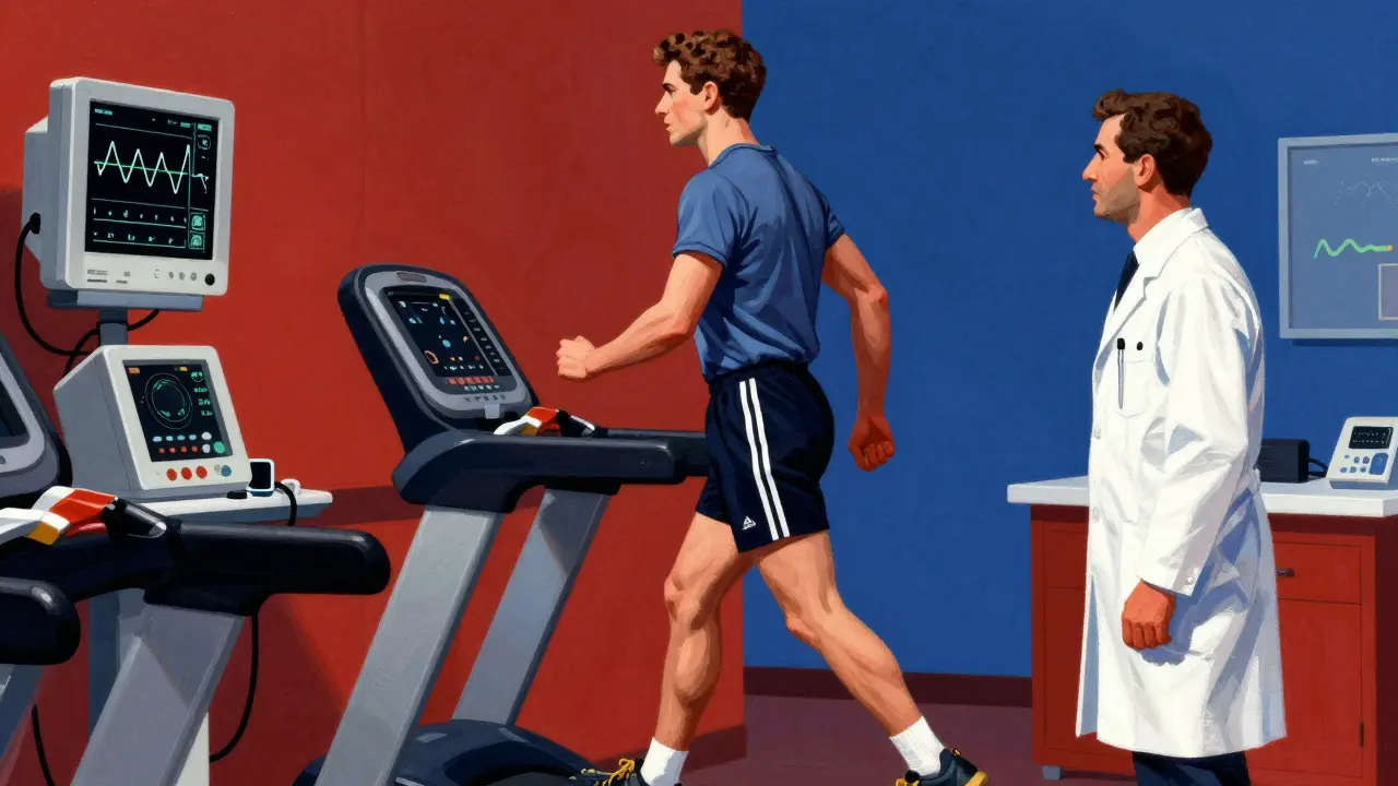
A stress test evaluates your heart's performance under exertion. It typically involves walking on a treadmill while connected to an ECG machine. The goal is to see if reduced blood flow causes symptoms or electrical changes that aren't visible at rest.
Which test is better?
Neither is inherently "better." An ECG is faster and cheaper but misses hidden blockages. A stress test provides more data on functional capacity but requires effort. Doctors choose based on your symptoms, risk factors, and ability to exercise.
How do I prepare for these tests?
For an ECG, no special prep is needed. For a stress test, avoid caffeine for 24 hours, wear comfortable shoes, and eat lightly beforehand. If you have arthritis or asthma, tell your doctor-you may need a chemical stress test instead.
Are stress tests safe?
Yes, they are very safe. Serious complications are rare. However, they are avoided in cases of recent heart attack, unstable arrhythmias, or severe heart failure. Medical staff monitor you closely throughout the procedure.
What if I can't walk on a treadmill?
You can undergo a chemical stress test. Medications like adenosine or dobutamine simulate exercise effects on the heart. This option is ideal for those with mobility issues, severe joint pain, or balance problems.
How accurate are these tests?
Standard ECG stress testing has about 68% sensitivity and 77% specificity for coronary artery disease. Adding imaging, like echocardiography or nuclear scans, improves accuracy significantly, especially for women who may experience false negatives on standard tests.
Do I need both tests?
Often, yes. An ECG is usually the first step. If results are inconclusive or symptoms persist, a stress test follows. Some patients get both on the same day, with the stress test incorporating continuous ECG monitoring.
How much do these tests cost?
Costs vary widely. In the US, heart CT scans average $404, stress echocardiograms around $514, and nuclear stress tests up to $946. Insurance coverage depends on medical necessity and provider networks.
What happens after the test?
Most facilities provide preliminary results immediately. You can usually resume normal activities right away unless told otherwise. Follow-up appointments discuss findings and next steps, such as medication adjustments or further imaging.
The Basics: How an ECG Works
An ECG is simple, fast, and painless. It records the electrical impulses that make your heart beat. Think of it as a photograph of your heart's rhythm at a single moment. Technicians place sticky electrodes on your skin-usually six on your chest, two on your wrists, and two on your ankles. These connect to a machine that translates electrical signals into wavy lines on paper or a screen.
The process takes only three to five minutes. Most of that time is spent attaching the electrodes correctly. Once connected, you lie still while the machine captures the data. The resulting graph shows P waves, QRS complexes, and T waves, each representing different phases of the heartbeat cycle. Doctors look for irregularities in timing, shape, or amplitude.
Why does this matter? Abnormal patterns can signal serious conditions. For example, ST-segment elevation might indicate an ongoing heart attack. Premature beats could suggest arrhythmia. Enlarged chambers may point to long-term high blood pressure damage. Because it's so quick, ECGs are often performed in emergency rooms, primary care offices, and even ambulances.
However, an ECG has limits. It only sees what's happening right now. If your heart acts normally at rest but struggles during activity, a resting ECG will miss it entirely. That's where stress tests come in.
Stress Tests: Pushing the Heart to Its Limits
A stress test forces your heart to work harder than usual. By increasing demand for oxygen-rich blood, doctors can spot blockages that hide when you're relaxed. There are two main types: exercise and chemical.
Exercise stress tests involve walking on a treadmill or pedaling a stationary bike. The intensity increases gradually, following protocols like the Bruce protocol. This starts slow-1.7 miles per hour with a 10% incline-and ramps up every three minutes until you reach your target heart rate or feel symptoms. Target heart rate is roughly 85% of your maximum predicted age (calculated as 220 minus your age).
During the test, technicians monitor four key metrics continuously:
- Heart rate (beats per minute)
- Blood pressure (mmHg)
- ECG waveform changes (millivolts)
- Oxygen saturation levels (%)
If you can't exercise due to arthritis, asthma, or recent surgery, pharmacological agents step in. Drugs like adenosine, dipyridamole, or dobutamine mimic exercise effects chemically. Adenosine widens blood vessels, forcing the heart to pump harder. Dobutamine increases heart rate and contractility directly. Chemical tests last longer-30 to 60 minutes-but offer similar diagnostic value.
Why go through all this trouble? Because many people have silent ischemia. Their arteries narrow enough to cause trouble during exertion but not at rest. Without a stress test, these issues remain undetected until a major event occurs.
Imaging Enhancements: Seeing Beyond Electrical Signals
Standard ECG stress tests rely solely on electrical readings. But adding imaging dramatically boosts accuracy. Two popular options exist: stress echocardiography and nuclear perfusion imaging.
Stress echocardiography combines ultrasound with exercise or chemical stress. Before and after stressing the heart, technicians capture images of its movement. Healthy muscle contracts strongly; damaged areas move weakly or paradoxically. This method avoids radiation exposure and offers superior specificity (88%) compared to nuclear methods (79%).
Nuclear stress testing uses radioactive tracers injected into your bloodstream. Cameras detect gamma rays emitted by isotopes like thallium-201 or technetium-99m. Areas receiving poor blood flow appear darker on the scan. Sensitivity reaches approximately 85%, higher than standard ECG alone. However, radiation exposure equals 9-12 millisieverts-comparable to three years of natural background radiation.
So which should you choose? Guidelines favor stress echocardiography for middle-aged women with intermediate risk due to better accuracy without radiation. Nuclear imaging suits complex cases requiring detailed anatomical maps. Always discuss pros and cons with your cardiologist.
Accuracy Matters: What Numbers Tell Us
No test is perfect. Understanding sensitivity and specificity helps set realistic expectations. Sensitivity measures how well a test detects true positives. Specificity gauges its ability to rule out false alarms.
According to meta-analyses covering over 24,000 patients, standard ECG stress testing achieves 68% sensitivity and 77% specificity for coronary artery disease. That means nearly one-third of actual blockages slip through unnoticed. False positives also occur, leading to unnecessary follow-ups.
Adding imaging closes this gap. Stress echocardiography raises specificity to 88%. Nuclear imaging pushes sensitivity toward 89%. Still, challenges persist. Women, especially premenopausal ones, face higher false-negative rates because of unique coronary microvascular physiology. Ethnic disparities also influence outcomes, underscoring the need for personalized approaches.
Prognostic markers matter too. Each extra minute spent exercising correlates with a 12% drop in future cardiac events. Functional capacity predicts survival better than almost any other factor measured during testing.
Preparing for Your Appointment
Proper preparation ensures reliable results. Start by avoiding caffeine for 24 hours before testing. Caffeine interferes with pharmacological agents used in chemical stress tests. Wear loose, comfortable clothing suitable for exercise. Bring supportive sneakers if doing an exercise test.
Eat lightly beforehand. Heavy meals divert blood flow away from muscles, skewing results. Stay hydrated but don't overdo fluids. Arrive early to complete paperwork and change into a gown if necessary.
Inform your team about medications, allergies, and medical history. Beta-blockers affect heart rate responses and may require temporary adjustment. Asthma sufferers should alert staff since certain drugs trigger bronchospasm.
Mental readiness counts too. Anxiety spikes cortisol levels, potentially affecting readings. Practice deep breathing techniques. Remember, millions undergo these tests safely every year. Trust the process.
Interpreting Results: What Comes Next?
After completion, wait times vary. Many clinics share preliminary findings before discharge. Full reports take days to weeks, depending on facility workload and complexity. Academic centers produce detailed documents averaging 3.2 pages with precise measurements. Community practices summarize essentials in shorter formats.
Inconclusive results happen frequently-about 25% of intermediate-risk patients need additional testing. Reasons include excessive motion artifacts, inadequate heart rate response, or ambiguous waveforms. Don't panic. Further evaluation clarifies uncertainties.
Treatment plans depend heavily on outcomes. Mild anomalies might warrant lifestyle modifications alone. Severe blockages could lead to angioplasty or bypass surgery. Medication adjustments address underlying hypertension, cholesterol, or diabetes contributing to vascular damage.
Follow-up schedules differ individually. High-risk individuals return sooner for repeat assessments. Low-risk folks enjoy extended intervals between visits. Regular monitoring maintains vigilance against progressing disease.
Future Directions: Technology Meets Tradition
Innovation transforms cardiovascular diagnostics rapidly. Artificial intelligence algorithms now interpret stress test data with 18-22% greater accuracy than traditional methods. Machine learning models analyze vast datasets identifying subtle patterns humans overlook.
Portable devices expand accessibility beyond hospital walls. FDA-cleared gadgets enable home-based screenings capturing comparable quality to institutional setups. Rural communities benefit immensely from decentralized care delivery models.
Speckle-tracking strain analysis enhances detection capabilities further. Combining this technique with stress echocardiography lifts sensitivity for microvascular dysfunction from 68% to 89% among symptomatic women lacking obstructive lesions. Precision medicine thrives on such advancements.
Despite emerging alternatives like coronary CT angiography gaining traction, stress testing retains relevance through 2030. Its versatility, affordability, and proven efficacy ensure continued adoption across diverse populations worldwide.